A Quantitative Analysis of Photoluminescence in North American Mammals
by Briana J. Roberson
Warnell School of Forestry and Natural Resources
Photoluminescence produced by excitation with ultraviolet light (UV) has been documented in an increasing number of nocturnal-crepuscular mammal species; however, little is currently known about quantitative characteristics of the trait. Here, I used museum specimens to examine spectral characteristics of UV-induced photoluminescence in 18 species of mammals representing a diversity of phylogenetic lineages. I observed varying blue, green/yellow, and pink/red photoluminescence among all species examined, and found genus and family level similarities in characteristics that diverge among higher taxonomic groupings. These findings contribute to the idea that photoluminescence is present in more mammal species than is currently documented. The results highlight the need for further quantitative exploration into the evolutionary and functional roles of photoluminescence across mammalian taxa.
ecology, museum specimens, luminescence, mammals
Introduction
Photoluminescence is the process by which luminophores (luminescent molecules) absorb excitation light at a shorter wavelength and produce an emission of light at a longer wavelength, and is well examined in many biological systems, especially plants and marine organisms. Chlorophyll-a fluorescence is extensively documented in both plants and phytoplankton (Harbinson, 2003). Photoluminescence is also present across the animal kingdom, with many recently published observations of mammals displaying the trait (Tumlison & Tumlison 2021; Reinhold et al 2023; Travouillon et al. 2023) suggesting it may be more widespread among vertebrates than originally thought.
While an increasing number of mammal species are observed to exhibit photoluminescence under ultraviolet light, there is much speculation as to whether photoluminescence may serve an explicit ecological function. Nearly all species in which luminescence has been observed are nocturnal, crepuscular, or fossorial (Kohler et. al 2019; Anich et. al 2021; Pynne et. al 2021), resulting in hypotheses for potential function in low-light environments. Understanding patterns between natural history, phylogeny, and characteristics of photoluminescence in order to investigate potential function is difficult, and links between observed characteristics in mammals are not easily isolated (Reinhold 2023). In addition, many studies have described emission characteristics, such as color, qualitatively (Pine et. al 1985; Kohler et. al 2019; Pynne et. al 2021; Tumlison & Tumlison, 2021; Reinhold et. al 2023), which further complicates comparative analyses. While a few studies have quantified spectra (Anich et. al 2021; Travouillon et. al 2023), they have focused on singular species or other regions not including North American species. Additional studies focusing on recording qualitative characteristics of photoluminescence in North American mammals would be valuable to better understand variation in the trait across taxa.
Examining color by wavelength of luminophore emission provides a way to quantify mammal photoluminescence, thereby creating opportunities for comparative analyses. Several luminophores have been identified as proximate causes for different colors of luminescence in mammal fur. Reddish UV fluorescence can be attributed to porphyrins across distantly related taxa from marsupials to placental mammals (Olson et. al 2021; Toussaint et. al 2023). It is speculated that blue UV fluorescence is produced as a result of tryptophan production (Nicholls & Reinits 1971; Reinhold et. al 2023). Similarity in peak absorption and emission spectra, and therefore color, has the potential to provide information on the source of photoluminescence either through shared luminophores across taxa or by the convergence of similar color by adaptive pressures.
Investigating relationships among sources of photoluminescence can provide guidance for consideration of evolutionary patterns. The trait may have appeared early in tetrapods, as fluorescence is widespread across multiple amphibian lineages (Lamb & Davis, 2020). If spectral characteristics are relatively consistent across species, the idea of fluorescence as a residual trait among tetrapods that some mammals have lost may be supported. If several species exhibit synonymous characteristics of emission, a shared physiological mechanism producing the emission may exist.
Here, I provide a quantitative analysis to confirm visual observations of UV-induced photoluminescence in eighteen species of mammals native to the southeastern United States. To do so, I examine the spectral characteristics of emission for differences within and between species and sex. Based on previous observations, I hypothesize that characteristics of emission differ between taxa but show greater similarities among more closely related species.
Methods
Specimens
I examined 18 species of mammals, 1-2 specimens per species (Appendix 1). Both sexes were examined for species with multiple specimens. All specimens were obtained from the Georgia Museum of Natural History (GMNH, Athens, Georgia) mammal collections.
Specimens were first visibly observed for presence of photoluminescence under 410 nm ultraviolet excitation light with a ±15 nm full width half maximum (FWHM) using yellow
UV-filtering lens (Circus NY, USA and LPSAFP, Amazon, China) to filter out excess UV and blue light. I photographed specimens in a dark box using a Nikon D5600 camera with an AF-P NIKKOR 18-55 mm lens (Nikon, Melville, New York, USA). For comparison, photographs were taken under only UV light and through the UV-blocking lens. In addition, I used a MIDOPT LP470 yellow longpass filter (Midwest Optical Systems Inc., Palatine, Illinois, USA) to filter out incident light under 470 nm to reduce visual noise and view photoluminescent emission color objectively without influence from ultraviolet and blue wavelengths. Specimen photos were digitized with a black background.
Scans
To record characteristics of photoluminescence, I quantified specimen emission spectra with an ILT950 spectroradiometer (International Light Technologies, Peabody, Massachusetts, USA) using an integration time of 4000 milliseconds. The probe was positioned at a height of 10mm over the area of brightest perceived photoluminescence. Intensity (brightness) of light was recorded as irradiance, measured in microwatts per square centimeter (µW/cm2). To account for confounding light in the dark room and to isolate photoluminescent emission from the excitation light, initial dark scans taken with no light present and reference scans taken of the excitation light were recorded before specimen scans, using a A253 Diffuse Reflectance Standard reference panel (International Light Technologies, Peabody, Massachusetts, USA). Dark scans and reference scans were subtracted from specimen scans using the following formula to ensure analyses only included light produced by specimens.
Photoluminescence = | Specimen irradiance values – (Dark scan irradiance values + reference irradiance values) |
I then processed scans to identify emission peaks using the dplyr package (version 1.1.1; Wickham, 2023) in R version 4.4.1 (R Core Team, 2024). I identified peak wavelength as the greatest irradiance value within each scan. Emission spectra for each species were graphed using the ‘ggplot’ package (version 3.3.3; Wickham, 2016).
Statistical Analysis
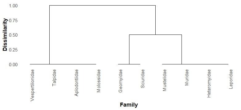
All analyses were conducted in R. I first used a Shapiro-Wilk test and found that peak wavelength was non-normally distributed. Therefore, to test for differences in peak wavelength within families, I used Kruskal-Wallis non-parametric tests for families with more than one species represented. Based on this information and examination of emission spectra, I decided to group species by family for cluster analysis. I created a dendrogram based on Jaccard dissimilarity for photoluminescent characteristics by categorizing emission peaks by color (blue: 450-515 nm, green/yellow: 515-600 nm, pink/red: 600-700 nm) using the ‘ape’ (Paradis et al. 2004), ‘ggdendro’ (de Vries & Ripley 2016), and ‘vegan’ (version 2.6-8; Oksanen 2010) packages. Data from six species of bats (Roberson et. al 2025) were included in the phylogenetic analysis.
Results
Visual Observations
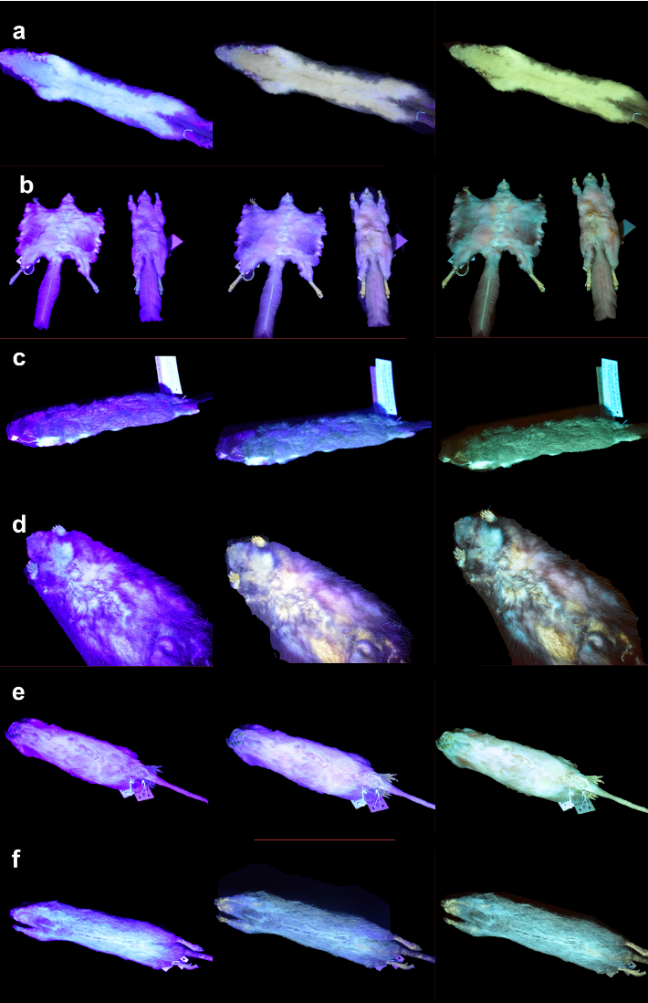
I observed photoluminescence of varying colors among the species examined (Figure 1). While differences in emission intensity were visually observed among specimens of the same species, color remained consistent. Using UV-filtering lenses to account for the high strength of blue light from the excitation source overwhelming the photoluminescence signal made a significant difference in the perceived color of specimens.
Blue photoluminescence was present on specimens of Kansas pocket gopher (G. arenarius), southeastern pocket gopher (G. pinetus), northern pocket gopher (T. talpoides), and Mexican spiny pocket mouse (H. irroratus). Yellow photoluminescence was specific to the mustelids, including short-tailed weasel (M. erminea) and long-tailed weasel (N. frenata). Pink color was observed on opossums (D. virginiana) and flying squirrels (G. volans). Minimal visually discernible color was observed in all other species examined.
Spectral Characteristics
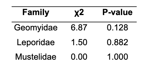
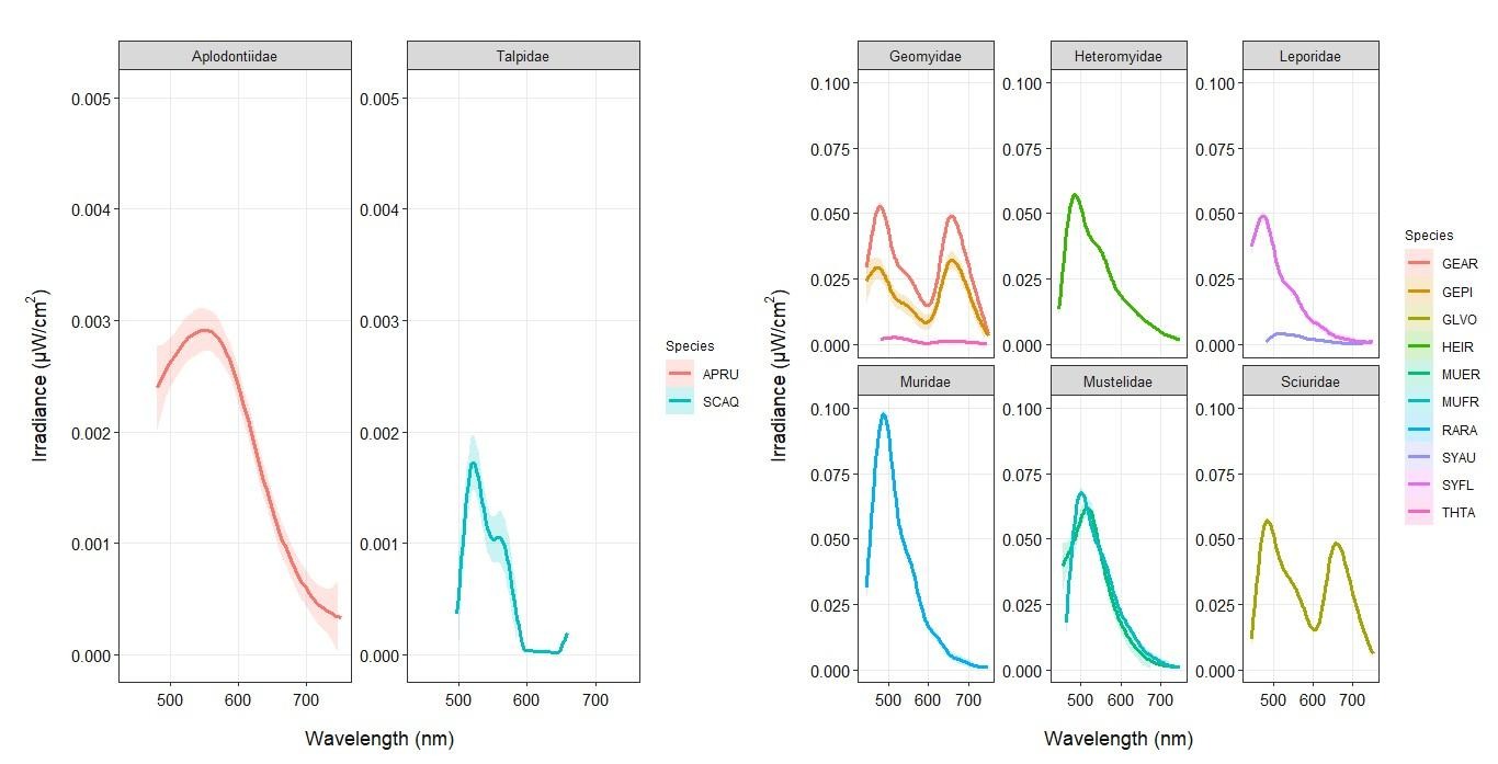
Photoluminescent emission was recorded in all species. Scans identified several peaks representing the range of color observed visually (Figure 1), including blue emission ranging from 456-511 nm, yellow/green emission ranging from 524-545 nm, and red/pink emission ranging from 621-690 nm (Figure 3). Several species exhibited lower irradiance levels, including A. rufa, S. aquaticus, and T. talpoides. I found no differences in peak wavelength within families (Table 1). Phylogenetic analyses indicated three groups: blue emission only (R. rattus, Sylvilagus, Heteromys, and Mustela), green emission only (all bat species, Scalopus, and Aplodontia) and simultaneous blue and red emission (Thomomys, Geomys, Glaucomys) (Figure 3).
Table 1. Results of Kruskal-Wallis tests comparing peak wavelength within family. Families with only one species examined were excluded.
Discussion
I provide observations of ultraviolet-induced photoluminescence in small mammal species of diverse taxa and life histories. These observations concur with other reviews of photoluminescence across mammal taxa, in that varying color and intensity of light were observed among species (Tumlison & Tumlison 2021; Reinhold et al. 2023; Travouillon et al. 2023). However, this analysis is the first to quantify these characteristics in North American species and provides a means to analyze phylogenetic patterns of photoluminescence across the taxa examined.
Based on these results, I suggest the presence of at least three individual luminophores responsible for the varying characteristics of photoluminescence observed. The blue emission I documented corroborates with spectral characteristics recorded in the platypus (Ornithorhynchus anatinus), koala (Phascolarctos cinereus), and Tasmanian devil (Sarcophilus harrisii) in Australia (Anich et. al 2021; Travouillon et al. 2023). In addition, the range of green photoluminescence found in bats (Reinhold 2022; Roberson et al. 2025), A. rufa, and S. aquaticus overlaps with the range of green emission documented in amphibians (Lamb & Davis 2021), and the multiple-peaked characteristics of emission observed in the Geomys genus, opossum, and flying squirrel (G. volans) are similar to the red fluorescence documented in the Pedetidae family (Olson et al. 2021), suggesting potential common luminophores among these species.
The existence of common luminophores across distantly related taxa suggests photoluminescence may be an ancestral trait in mammals. While I observed similar characteristics within families, emission peaks did not appear to follow phylogenetic patterns at the order level, with many species within Rodentia exhibiting differing characteristics of emission (Figure 3). The similarity of emission within families combined with the variation in persistence of luminophores among higher taxonomic groups suggests adaptive pressures specific to life history may have modified the trait to produce the diversity of characteristics present in modern mammals. Blue emission was present in all members of Rodentia except for A. rufa (Figure 2), suggesting that blue photoluminescence currently thought to originate from tryptophan metabolites (Nicholls & Reinits 1971; Reinhold 2023) may be a common trait among rodents that has been lost in Aplodontiidae. Green photoluminescence observed in bats (Reinhold 2022; Roberson et al. 2025) appears primarily within the skin, and I detected a peak within the same wavelength range in other mammal species that was not observed visually, so it is possible that the luminophore responsible for green fluorescence is cutaneous. Lamb & Davis (2021) found widespread cutaneous green photoluminescence in amphibians. Accordingly, there is the potential for green photoluminescence to have originated in an early shared ancestor. Additional research relating characteristics of emission to phylogenetic patterns and additional examinations of ranging mammal, amphibian, and reptile taxa would be required to gain more insight into the evolution of specific luminophores.
While there were no differences in color observed within the same family, there were differences in irradiance levels with several species (A. rufa, S. aquaticus, and T. talpoides) exhibiting lower irradiance values. This suggests the potential for the same luminophores to be present in greater amounts within some species, and less in others. Adaptive pressures related to life history could account for this difference. For example, these three species are all primarily fossorial (dwelling underground) and are less likely to be exposed to nighttime ultraviolet light than other nocturnal mammals. Additional testing with a larger number of specimens and a consideration of optical color reception among the species examined would be required to understand whether the colors and levels of irradiance documented are representative of species and truly visually relevant. In addition, testing at variable excitation wavelengths would differentiate whether emission is due to the photoluminescent mechanisms of true fluorescence or light scattering (Shinde et al. 2012).
These results suggest a shared origin of photoluminescence within species and between closely related species, but a phylogenetic divergence among higher taxonomic groups. Additional analysis of within-species replicates and comparisons of closely related species would be necessary to correlate the observed divergence to adaptive pressures relating to ecology and/or life history. Investigation of phylogenetic patterns through genetics, specifically genome wide association studies (GWAS), may be valuable in identifying specific genetic variations associated with observed spectral characteristics or luminophore presence and would allow better understanding of evolutionary patterns and history of the trait. I suggest future studies combine multiple approaches with quantification of photoluminescence characteristics to explicitly examine evolutionary history and potential ecological functions.
References
Anich, P. S., S. Anthony, M. Carlson, A. Gunnelson, A. M. Kohler, J. G. Martin, and E. R. Olson. 2021. Biofluorescence in the platypus (Ornithorhynchus anatinus). Mammalia 85:179–181.
Clarke, R. J., and A. Oprysa. 2004. Fluorescence and Light Scattering. Journal of Chemical Education 81:705-707.
Kohler, A. M., E. R. Olson, J. G. Martin, and P. S. Anich. 2019. Ultraviolet fluorescence discovered in New World flying squirrels (Glaucomys). Journal of Mammalogy 100:21–30.
Lagorio, M. G., G. B. Cordon, and A. Iriel. 2015. Reviewing the relevance of fluorescence in biological systems. Photochemical & Photobiological Sciences 14:1538–1559.
Lamb, J. Y., and M. P. Davis. 2020. Salamanders and other amphibians are aglow with biofluorescence. Scientific Reports 10:2821.
Nicholls, E. M., and K. G. Rienits. 1971. Tryptophan derivatives and pigment in the hair of some australian marsupials. International Journal of Biochemistry 2:593–603.
Oksanen, J., F. G. Blanchet, M. Friendly, R. Kindt, P. Legendre, D. McGlinn, P. R. Minchin, R. B. O’Hara, G. L. Simpson, P. Solymos, M. H. H. Stevens, E. Szoecs, and H.
Wagner. 2022. vegan: Community Ecology Package. R package version 2.6-4.
Olson, E. R., M. R. Carlson, V. M. S. Ramanujam, L. Sears, S. E. Anthony, P. S. Anich, L. Ramon, A. Hulstrand, M. Jurewicz, A. S. Gunnelson, A. M. Kohler, and J. G. Martin. 2021. Vivid biofluorescence discovered in the nocturnal Springhare (Pedetidae). Scientific Reports 11:4125.
Paradis, E., J. Claude, and K. Strimmer. 2004. APE: Analyses of Phylogenetics and Evolution in R language. Bioinformatics 20:289–290.
Pine, R. H., J. E. Rice, J. E. Bucher, D. H. Jr. Tank, and A. M. Greenhall. 1985. Labile pigments and fluorescent pelage in didelphid marsupials. Mammalia 49.
Pynne, J. T., S. B. Castleberry, L. M. Conner, C. W. Piper, E. I. Parsons, R. A. Gitzen, S. I. Duncan, J. D. Austin, and R. A. McCleery. 2021. Ultraviolet biofluorescence in pocket gophers. The American Midland Naturalist 186.
Reinhold, L. 2022. Photoluminescent yellow wing markings of Eastern Tube-nosed Fruit Bats (Nyctimene robinsoni). North Queensland Naturalist 52:69-74.
Reinhold, L. 2023. Widespread fluorescence in the fur of mammals of the Australian wet tropics. Proceedings of the Royal Society of Queensland 132.
Reinhold, L. M., T. L. Rymer, K. M. Helgen, and D. T. Wilson. 2023. Photoluminescence in mammal fur: 111 years of research. Journal of Mammalogy 104:892–906.
R Core Team. 2023. R: A language and environment for statistical computing. R Foundation for Statistical Computing, Vienna, Austria. URL: https://www.R-project.org/
Roberson, B. J., Perea, S., D. DeRose‐Broeckert, and S. B. Castleberry. 2025. Glowing green: A quantitative analysis of photoluminescence in six North American bat species. Ecology and Evolution 15:e71885.
Shinde, K. N., S. J. Dhoble, H. C. Swart, and K. Park. 2012. Phosphate phosphors for solid-state lighting. Springer Berlin Heidelberg, Berlin, Heidelberg.
Toussaint, S. L. D., J. Ponstein, M. Thoury, R. Métivier, D. C. Kalthoff, B. Habermeyer, R. Guilard, S. Bock, P. Mortensen, S. Sandberg, P. Gueriau, and E. Amson. 2023. Fur glowing under ultraviolet: in situ analysis of porphyrin accumulation in the skin appendages of mammals. Integrative Zoology 18:15–26.
Travouillon, K. J., C. Cooper, J. T. Bouzin, L. S. Umbrello, and S. W. Lewis. 2023. All-a-glow: Spectral characteristics confirm widespread fluorescence for mammals. Royal Society Open Science 10:230325.
Tumlison, C. R., and T. Tumlison. 2021. Investigation of fluorescence in selected mammals of Arkansas. Journal of the Arkansas Academy of Science 75.
Willis, C. K. R., and R. M. Brigham. 2007. Social thermoregulation exerts more influence than microclimate on forest roost preferences by a cavity-dwelling bat. Behavioral Ecology and Sociobiology 62:97–108.
With, T. K. 1978. On porphyrins in feathers of owls and bustards. International Journal of Biochemistry 9:893–895.
Zeileis, A., and G. Grothendieck. 2005. Zoo: An S3 package for time series analysis. Journal of Statistical Software 14:1-27.
Acknowledgments: I thank Dr. Steven B. Castleberry for his excellent mentorship, Nicole Pontzer at the Georgia Museum of Natural History for providing specimens, Daniel DeRose-Broeckert for support with data collection, and Dr. Billy Hammond at the University of Georgia Vision Sciences laboratory for providing access to the lab and equipment.
Citation Style: Ecology
Appendix 1. List of species examined
| Species | Scientific Name |
| Mountain Beaver | Aplodontia rufa |
| Kansas Pocket Gopher | Geomys arenarius |
| Southeastern Pocket Gopher | Geomys pinetus |
| Flying Squirrel | Glaucomys volans |
| Mexican Spiny Pocket Mouse | Heteromys irroratus |
| Short-tailed Weasel | Mustela erminea |
| Long-tailed Weasel | Neogale frenata |
| Black Rat | Rattus rattus |
| Eastern Mole | Scalopus aquaticus |
| Desert Cottontail | Sylvilagus auduboni |
| Eastern Cottontail | Sylvilagus floridanus |
| Northern Pocket Gopher | Thomomys talpoides |
| Big Brown Bat | Eptesicus fuscus |
| Eastern Red Bat | Lasiurus borealis |
| Seminole Bat | Lasiurus seminolus |
| Southeastern Myotis | Myotis austroriparius |
| Gray Bat | Myotis grisescens |
| Brazilian Free-tailed Bat | Tadarida brasiliensis |